Thursday, December 16, 2010

The Dry Eye

A patient might complain of dryness of the eyes simply because the conjunctiva is inflamed, but when the tear film really is defective, the patient might complain of soreness and irritation rather than dryness. The diagnosis of a dry eye depends on a careful examination and it is quite erroneous to assume that the tear film is inadequate simply because the patient complains of dryness, or even if the symptoms appear to be improved by artificial tears.

The normal tear film consists of three layers and the integrity of this film is essential for comfort and more importantly for good vision. The anterior, or outermost, layer is formed by the oily secretion of the meibomian glands and the layer next to the cornea is mucinous to allow proper wetting by the watery component of the tears, which lies sandwiched between the two. This three-layered film is constantly maintained by the act of blinking.

Causes

• Systemic disease with lacrimal gland
involvement:
– sarcoidosis
– rheumatoid arthritis (Sjögren’s
syndrome).
• Trachoma (chlamydial conjunctivitis and
keratitis – see next chapter).
• Neuroparalytic keratitis.
• Exposure keratitis.
• Old age.
• Other rare causes.

Signs

Slit-lamp Examination
In a normal subject, the tear film is evident as a rim of fluid along the lid margin and a deficiency of this can be seen by direct examination. Prolonged deficiency of tears can be associated with the presence of filaments – microscopic strands of mucus and epithelial cells, which stain with Rose Bengal. Punctate staining of the corneal epithelium is also seen after applying a drop of fluorescein. In some dry eye syndromes, for example, ocular pemphigoid and Stevens–Johnson syndrome, keratinisation of the cornea and conjunctiva with the formation of contracting adhesions between the opposed surfaces of the conjunctiva occurs. A similar change is apparent following chemical or thermal burns of the eyes.
Schirmer’s Test
One end of a special filter paper strip is placed between the globe and the lower eyelid. The other end projects forward and the time taken for the tears to wet the projecting strip is measured. The test is not an accurate measure of tear secretion but it provides a useful guide
Tear Film Break-up Time
Using the slit-lamp microscope, the time for the tear film to break up when the patient stops blinking is measured. This test is sometimes used as an index of mucin deficiency.

Management of the Dry Eye

This, of course, depends on the cause of the dry eye and the underlying systemic cause might require treatment in the first place. Artificial tear drops are a mainstay in treatment and various types are available, their use depending on which component of the tear film is defective. In severe cases, it might be necessary to consider temporary or permanent occlusion of the lacrimal puncta.


Blunt Trauma Or Injury To The Eye

The fact that injuries to the eye and its surrounding region demand special attention and create great concern for patient and doctor is self-evident when the eye alone is involved, but when other life-threatening injuries are present, the eye injury, seeming slight at the time, might be overlooked. Sometimes, the eyelids might be so swollen that it is difficult to examine the eyes and a serious perforating injury could be obscured.When other injuries are present and an anaesthetic is needed, it is essential that the eyes are examined carefully, if possible under
the same anaesthetic. As in the case of injuries elsewhere, those to the eye demand urgent and immediate treatment, and neglect can result in tragedy even though the problem might have at first seemed slight.

Injuries to the Globe


Contusion


The eye casualty officer comes to recognize a familiar pattern of contusion, the effect of squash ball injuries and blows from flying objects in industry or after criminal assault. The extent of damage to the eye from contusion depends on whether it has been possible to close the eyelids in time before the moment of impact. If the lids have been closed, bruising and swelling of the eyelids is marked and the injury to the eye might be minimal. The eyes themselves must always be
carefully examined, even when there is extreme swelling of the lids. It is always possible to examine an eye, if necessary using an eye speculum under general anaesthesia.The important clinical features of contusion injury are best considered by looking at the anatomical parts of the eye.

Cornea


The most common injury to the cornea is the corneal abrasion. Here, the patient
experiences a sharp pain in the eye in the early morning usually on waking, sometimes many months after the initial injury. The cornea is to be examined carefully with the slitlamp biomicroscope. This problem of recurrence is a reason to treat these abrasions with some care and to provide the patient with a lubricating
ointment to be used at night for some time after the original injury has healed. Sometimes, recurrent abrasion results from a rare inherited disorder of the corneal epithelium. When a patient presents with a corneal abrasion, the eyelids are often swollen perhaps from rubbing and the distress and agitation can be considerable. Examination may be impossible without first instilling a drop of local anaesthetic.
These drops should never be continued as treatment because they could seriously delay the healing of the cornea.

Anterior Chamber


A small bleed into the anterior chamber of the eye is seen as a fluid level of blood inferiorly (“hyphaema”). This is a sign of potential problems because of the risk of secondary bleeding after two or three days. This risk is especially serious in children and the complication can lead to secondary glaucoma and at worst, the loss of the eye.The parents need to be warned about this if there is a hyphaema. Treatment is by strict rest with little or no head movement to avoid further bleeding and regular measurement of the intraocular pressure.

Iris

When confronted by a flying missile, the normal reaction is to attempt to close the eyelids and to rotate the eyes upward. This is the reason why the commonest point of impact is the lower temporal part of the eye and it is in this region of the iris that one is most likely to see peripheral iris tears (“iridodialysis”). When the eye is compressed the iris periphery is torn at its root, leaving a crescentic gap, which looks black, but through which the fundus and red reflex can be observed. Such an injury also provides an excellent view of the peripheral part of the lens and the zonular ligament
Contusion can result not in a tear of the iris root, but in a tangential splitting of the iris and ciliary body from the sclera producing recession of the angle of the anterior chamber; the appearance is often associated with secondary glaucoma, sometimes many years after the injury and is identified using the special contact lens known as the gonioscope. A sudden impact on the eye can also produce microscopic radial tears in the pupillary  sphincter of the iris. This could be a subtle microscopic sign of previous injury when no other signs are present, or the damage might be more severe, resulting in persistent dilatation of the pupil (traumatic mydriasis). Unless the eye is examined, this widening of the pupil after injury can be mistaken for a third cranial nerve palsy.

Lens

Any severe contusion of the eye is liable to cause traumatic cataract(in rossette shaped pattern), but the lens might not become opaque for many years after the injury. The lens can also become subluxated (slightly displaced because of partial rupture of the zonular ligament) or even dislocated either anteriorly into the anterior chamber or posteriorly into the vitreous.

Vitreous


The vitreous can become displaced from its attachments around the processes of the ciliary body or around the optic disc after a contusion injury if it has not already undergone this change as part of the normal ageing process. The patient might be aware of something floating in front of the vision. More extensive floating black spots can indicate a vitreous haemorrhage caused by excessive vitreous traction on a retinal blood vessel. Although such haemorrhages usually clear completely in time, they tend to accompany more serious damage to the retina,which can only be fully revealed once clearing has taken place.

Retina


Bruising and oedema of the retina are seen as grey areas with scattered haemorrhages. The macular region is susceptible to oedema after contusion injuries, causing permanent damage to the reading vision. Just as tears can occur to
the peripheral iris, so a similar problem is seen in the peripheral retina. These crescent-shaped retinal dialyses are also most common in the lower temporal quadrant and their importance lies in the fact that they may lead to a detachment of the retina unless the tear is sealed by laser treatment. Any significant contusion injury of the eye requires a careful inspection of the peripheral retina.

Choroid

Tears in the choroid following contusion have a characteristic appearance. They are concentric with the disc and are seen as white crescents where the sclera is exposed. When near the macula, there is usually permanent damage to the central vision. They are also potential sites for choroidal neovascularisation.

Optic Nerve

A variable degree of optic atrophy can become apparent a few weeks after a contusion injury. Blunt injuries to the eye can cause bleeding into the optic nerve sheath or tearing of the tiny pial blood vessels that supply the nerve, both resulting in complete, irreversible loss of vision on the affected side. Attempts have been made to relieve the situation by emergency decompression of the optic nerve, nerve sheath fenestration, use of hyperbaric oxygen and highdose steroids. No treatment has shown a clear benefit except optic nerve decompression in specific circumstances.

Perforation

As soon as the globe of the eye is penetrated, there is a serious risk of infection. The vitreous is an excellent culture medium and in the pre-antibiotic era, eyes were totally lost within two or three days as a result of this. A perforating wound of the eye must, therefore, be considered a surgical emergency. Perforating injuries are seen in children from scissor blades, screwdrivers, darts and other more bizarre objects. In adults, there has been a dramatic fall in the incidence of such injuries since the introduction of compulsory seat belts but “do-it-yourself” accidents and assaults still take their toll. Following such an injury it is important to consider the possibility of an intraocular foreign body, especially when there is a history of using a hammer and chisel. The outcome of a perforating injury is dependent on the depth of penetration and the care with which the wound is cleaned and sutured. If the cornea alone is damaged, excellent results can be obtained by careful suturing under general anaesthesia using the operating microscope. If the lens has been damaged, early cataract surgery might be needed and deeper penetration can result in the need for retinal detachment surgery. On admission or in the casualty department, the patient is given tetanus prophylaxis and both systemic and local antibiotics. If early surgery under general anaesthesia is likely to be needed, it is better for the patient not to eat or drink to avoid delays in hospital. If it becomes clear that the injury is a serious one, it is better to warn the patient at an early stage about the possible risk of losing the sight of the eye or even the need to replace it with an artificial one.

Intraocular Foreign Body

Metallic foreign bodies tend to enter the eyes of those who operate high-speed grinders without goggles or those using a hammer and chisel on metal without eye protection. These injuries might seem slight at first and sometimes patients do not attach much importance to them.Any such eye injury with this occupational history warrants an X-ray of the eye. When ferrous metals remain in the eye they can cause immediate infection, or at a later date the deposition of ferrous salts, in a process known as siderosis. This can eventually lead to blindness of the eye. Other metals also tend to give reactions, particularly copper and for this reason the metallic fragment should be removed. This is achieved either by using intravitreous forceps under microscopic control or using a magnet. The exact surgical technique is planned beforehand once the foreign body has been accurately localised in the eye. Airgun pellets cause particularly severe eye injuries and the eye is often lost because of the extensive disruption at the time of the injury. Some intraocular foreign bodies, such as glass particles or some alloys, might be tolerated quite well and a decision could have to be made as to whether observation is preferable in the first instance. This especially applies when the sight of the eye remains good.When a foreign body is not to be removed immediately, many ophthalmologists would insert intravitreal antibiotics as a prophylactic measure against endophthalmitis. When a foreign body is found lying deeply in the cornea, its removal can result in loss of aqueous and collapse of the anterior chamber. It is prudent to arrange that removal should be done under full sterile conditions in the operating theatre, where the corneal wound can be sutured if necessary.

Sympathetic Ophthalmia


This rare complication of perforation is more common in children. The injured eye remains markedly inflamed and the wound might have been cleaned inadequately or too late. Over a period of two weeks to several months or even years a particular type of inflammatory response begins in the uvea and subsequently a similar reaction occurs in the other eye. The inflammation in both eyes can be so severe as to cause blindness. The condition does, however,  respond well to steroid treatment and it is extremely rare. Occasionally, one sees patients who have an artificial eye complaining of transient blurring of the vision of their remaining eye. They need to be examined carefully for signs of uveitis.

Injuries to the Eyelids


Loss or destruction of eyelid tissue should always be treated as a threat to vision. The upper lid especially is important in this respect. The immediate concern is to ensure that the cornea is properly covered when the eyelids are closed. If more than one-third of the margin of the upper lid is lost, this must be replaced by grafting from the lower lid.When less than one-third is missing, the gaping wound can usually be closed directly. Up to one-third of the lower lid can also be closed by direct suturing. When more than this is lost or when it has been transferred to the upper lid, a slide of tissue from the lateral canthus can be effected, combined if necessary with a rotating cheek flap. One of the most important features of the repair of lid injuries is the method of suturing. If the lid margin is involved, the repair should be made using the operating microscope and the fine suture material available in an eye department. An untidy repair can result in a permanently watering eye because of kinking of the eyelid. This interferes with the proper moistening of the cornea during blinking or when asleep. Special attention must be
paid when the medial part of the eyelid has been torn, as this contains the lacrimal canaliculus. Again, unless repair is carried out using an accurate technique under general anaesthesia in theatre, the risk of a permanently watering eye is increased.

Injuries to the Orbit

The most common type is the “blow-out fracture”. Here the globe and contents of the orbit are forced backwards, causing fracture of the orbital floor and displacement of bone downwards into the antrum of the maxillary sinus. The inferior rectus muscle becomes tethered in the wound so that there is mechanical limitation of upward movement. The infraorbital nerve, which traverses the orbital floor, can also be injured, producing anaesthesia of the skin of the cheek. Once the surrounding swelling has subsided, the posterior displacement of the globe becomes
obvious and the globe of the eye itself often shows evidence of contusion. A considerable improvement from the functional and cosmetic point of view can be obtained by positioning a plastic or Teflon implant in the floor of the orbit after freeing the prolapsed tissue. Fractures of the skull that extend into the orbit can be accompanied by retro-orbital haemorrhage and proptosis.Cranial nerve palsies affecting the ocular movements are also commonly seen in this type of injury and the vision can be affected by optic nerve damage.A blow on the eye can result in sudden blindness with at first no other evidence of injury (apart from an afferent pupillary defect), but subsequently, the optic disc becomes pale and atrophic after two or three weeks.


Progressive Myopia

Progressive myopia, also known as degenerative myopia or malignant myopia or pathological myopia, is a clinical type of myopia or short-sightedness associated with some typical degenerative changes in the fundus.

Associations: Pathological myopia has been associated with other ocular and systemic diseases. These conditions include Down’s Syndrome, Ocular Albinism, Infantile Glaucoma, Marfan’s Syndrome, Retinopathy of Prematurity, Ehler’s-Danlos Syndrome, low birth weight, and maternal alcoholism.

Symptoms: Impaired distant vision, eye-strain, black floaters and flashes of light, delayed dark adaptation or even night blindness, exophoria or divergent squint.

Signs: 1.prominent eyeball(pseudo-proptosis),2.larger cornea, 3.large pupil, 4.deep anterior chamber, 5.degeneration of vitreous.

Ophthalmoscopically, 1.Optic disc-large disc with mild pallor, temporal or annular crescent, posterior staphyloma. 2.Macula: dull, Foster-Fuchs fleck(spot) & degeneration. 3.General fundus: pale and tessellated appearance. Choroidal sclerosis and patches of choroidal atrophy. 4.Peripheral retina: Cystoid degeneration

Complications: 1.Retinal tear->Vitreous Haemorrhage 2.Retinal Detachment 3.Degeneration of vitreous 4.Primary Open Angle Glaucoma 5.Posterior cortical cataract.

Treatment:

1.Glasses: Appropriate concave lens with slight under-correction (Disadvantage: minification of object, image distortion, decrease in field of vision, cosmetic problem)

2.Contact lenses: (Advantage:less minification of image, no image distortion, no decrease in field of vision, no cosmetic problem)

3.Surgery: i.Radial keratotomy, ii.Keratomileusis iii.Fukala’s operation iv.Clear lens extraction(ECCE with PC-IOL) v.Minus phakic AC or PC IOL implantation vi.Intra-corneal rings insertion in paracentral area of cornea

4.LASER:1.Photo-refractive keratoplasty. 2.LASIK(Laser Assisted Stromal In Situ Keratomileusis)-more popular, but costly.

D.General Measures: Nutritious diet, proper positioning of head while reading in bright illumination, Fresh environment.


After Cataract or Posterior Capsule Opacification

Posterior Capsule Opacification, also called After Cataract is the most common long-term complication of Extra Capsular Cataract Extraction.

Posterior capsular opacification can occur months or years after cataract surgery, unlike most other cataract surgery complications that tend to occur during or soon after the procedure. Within 2 to 5 years after surgery, PCO causes decreased visual acuity in 20 percent to 40 percent of eyes that have had cataract surgery.

Risk Factors: The problem is age-dependent and occurs more frequently in young patients. The rate of PCO among children who have surgery to remove congenital cataracts ranges from 20%-50% by 2-4 years. The problem occurs more frequently in patients with PMMA lens and hydrophilic acrylic lenses.

Pathogenesis: Opacification of the posterior capsule appears to be influenced by lens epithelial cells that are left behind in the eye during cataract removal. These cells multiply, migrate across the posterior lens capsule and undergo changes that cause fibrous or pearl-type opacities in the capsule. The exact mechanism is not completely understood, but it appears to be a aberrant would healing response to cataract surgery.

Treatment: Currently, the only treatment of PCO is a type of laser surgery called Nd:YAG laser capsulotomy. In this procedure, a laser is used to create a central opening in the cloudy posterior capsule to restore sight. The procedure is quick and easy, but possible complications of laser capsulotomy include retinal detachment, damage to the intraocular lens (IOL), increase in intraocular pressure and other problems.


Wednesday, December 15, 2010

Chalazion: Symptoms, Treatment, Complications

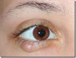
chalazion Chalazion, also known as a meibomian gland lipogranuloma, is a cyst in the eyelid that is caused by inflammation of a blocked meibomian gland, usually on the upper eyelid. Chalazia differ from styes (hordeola) in that they are subacute, nontender, and usually painless nodules. They may become acutely inflamed but, unlike a stye, chalazia usually point inside the lid rather than on the lid margin.
Signs and symptoms

  • Swelling on the eyelid
  • Eyelid tenderness
  • Sensitivity to light
  • Increased tearing
  • Heaviness of the eyelid

Treatment:

Topical antibiotic eye drops or ointment (e.g. chloramphenicol or fusidic acid) are sometimes used for the initial acute infection, but are otherwise of little value in treating a chalazion. Chalazia will often disappear without further treatment within a few months and virtually all will resorb within two years.

If they continue to enlarge or fail to settle within a few months, then smaller lesions may be injected with a corticosteroid or larger ones may be surgically removed using local anesthesia.

Chalazion Surgery:

Chalazion removal surgery is performed under local or general anesthesia. Commonly, general anesthesia is administered in children to make sure they stay still and no injury to the eye occurs. Local anesthesia is used in adults and it is applied with a small injection into the eyelid. The discomfort of the injection is minimized with the help of an anesthetic cream which is applied locally.

The chalazion may be removed in two ways, depending on the size of cyst. Relatively small chalazia are removed through a small cut at the back of the eyelid. The surgeon lifts the eyelid so he can have access to the back of its surface and makes an incision of approximately 3mm just on top of the chalazion. The lump is then removed and pressure is applied for a few minutes to stop any oozing of blood that may occur because of the operation. Surgery of small chalazia does not require stitches as the cut is at the back of the eyelid and therefore the cut cannot be seen and the cosmetic result is excellent.

Larger chalazia are removed through an incision in front of the eyelid. Larger chalazia usually push on the skin of the eyelid and this is the main reason why doctors prefer removing them this way. The cut is not larger than 3 mm and it is performed on top of the chalazion. The lump is removed and then pressure is applied on the incision so oozing is prevented. This type of surgery is closed with very fine stitches. They are hardly visible and they are usually removed within a week after the surgery has been performed. Although chalazia are rarely dangerous, every removed chalazion is sent to the laboratory to be examined under a microscope because very rarely it can harbor cancer.

When surgery for chalazion is considered, patients who take aspirin or any medication that contains aspirin are advised to stop taking them one week before the procedure as they may cause bleeding. There are several tests taken prior the surgery to make sure the patient is in good condition for the operation.

In rare cases, patients are kept overnight in the hospital after chalazion surgery. These include cases in which complications occurred and the patient needs to be closely monitored. In most cases however, patients are able to go home after the operation has ended.

The recovery process is easy and quite fast. Most patients experience some very minor discomfort in the eye which can be easily controlled by taking painkilling medication. Patients are however recommended to avoid getting water in the eye for up to 10 days after surgery, they may wash, bathe or shower but they must be careful in keeping the area dry and clean. Makeup may be worn after one month after surgery. Patients who wear contact lenses are recommended to not wear on in the operated side for at least eight weeks to prevent infection and potential complications.

Commonly, patients receive eye drops to prevent infection and swellings in the eye and pain medication that will help them cope with the pain and discomfort in the eyelid and eye. One can use paracetamol rather than aspirin to control the pain. Also, after surgery, a pad and protective plastic shield are used to apply pressure on the eye in order to prevent leakage of blood after the operation and which may be removed 6 to 8 hours after the procedure.

People who underwent chalazion surgery are normally asked to check up their operation three to four weeks after surgery has been performed. They may start driving the day after surgery and they may get back to work in one or two days.

Chalazion surgery is a safe procedure and complications occur very seldom. Serious complications that require another operation to be fixed are also very rare. Among potential complications, although rare, there is infection, bleeding or the recurrence of the chalazion.

Complications:

A large chalazion can cause astigmatism due to pressure on the cornea.

As laser eye surgery involves shaping the cornea by burning parts of it away, weakening its structure, post operation patients can be left predisposed to deformation of the cornea from small chalazia.

Complications including, but not limited to hypopigmentation may occur with corticosteroid injection.

Recurring chalazia in the same area may sometimes be a symptom of sebaceous cell carcinoma, albeit rarely. This is a type of cancer.

Sometimes, as a last resort, surgery is performed. The eyelid is injected with a local anesthetic, a clamp is put on the eyelid, then the eyelid is turned over, an incision is made on the inside of the eyelid, and the chalazion is drained and scraped out. A scar on the upper lid can cause discomfort as some patients feel the scar as they blink. Of course as surgeries are intrusive and damage healthy tissue (e.g. leaving behind scar tissue or possibly even causing blepharitis), given other options, less intrusive treatment is always preferable. Similarly, chalazia may recur once the eye is predisposed and surgical intervention each time is not possible. So surgery should be considered only as a last resort, performed on as few as 5% of all chalazia patients.


Retinitis Pigmentosa: Signs, Diagnosis And Treatment

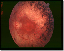
retinitis pigmentosa Retinitis pigmentosa (RP) is a group of inherited disorders characterized by progressive peripheral vision loss and night vision difficulties (nyctalopia) that can lead to central vision loss.

Signs:

Mottling of the retinal pigment epithelium with black bone-spicule pigmentation is typically indicative (or pathognomonic) of retinitis pigmentosa. Other ocular features include waxy pallor of the optic nerve head, attenuation (thinning) of the retinal vessels, cellophane maculopathy, cystic macular edema and posterior subcapsular cataract.

Diagnosis:

The diagnosis of retinitis pigmentosa relies upon documentation of progressive loss in photoreceptor cell function by electroretinography (ERG) and visual field testing.

The mode of inheritance of RP is determined by family history. At least 35 different genes or loci are known to cause "nonsyndromic RP" (RP that is not the result of another disease or part of a wider syndrome).

DNA testing is available on a clinical basis for:

  • RLBP1 (autosomal recessive, Bothnia type RP)
  • RP1 (autosomal dominant, RP1)
  • RHO (autosomal dominant, RP4)
  • RDS (autosomal dominant, RP7)
  • PRPF8 (autosomal dominant, RP13)
  • PRPF3 (autosomal dominant, RP18)
  • CRB1 (autosomal recessive, RP12)
  • ABCA4 (autosomal recessive, RP19)
  • RPE65 (autosomal recessive, RP20)

Treatment:

Although incurable, the progression of the disease can be reduced by the daily intake of 15000 IU (equivalent to 4.5 mg) of vitamin A palmitate. Recent studies have shown that proper vitamin A supplementation can postpone blindness by up to 10 years (by reducing the 10% loss pa to 8.3% pa),

Associations:

Retinitis pigmentosa is seen in a variety of diseases, so the differential of this sign alone, is broad.

  • RP combined with deafness (congenital or progressive) is called Usher syndrome.
  • RP combined with opthalmoplegia, dysphagia, ataxia, and cardiac conduction defects is seen in the mitochondrial DNA disorder Kearns-Sayre syndrome (aka Ragged Red Fiber Myopathy)
  • RP combined with retardation, peripheral neuropathy, acanthotic (spiked) RBCs, ataxia, steatorrhea, is absence of VLDL is seen in abetalipoproteinemia.


Classification of Corneal Ulcers On The Basis Of Aetiology

Microbial ulcers
These are caused by Staph or Strep species, present with pain, redness, lid
oedema and discharge that varies according to the duration and severity of the
ulcer. The ulcer is well circumscribed and may be central or at mid-periphery of
the cornea. The borders of the ulcer are usually round or oval. Stromal infiltration
may be present under the base of the ulcer with a yellowish white appearance.
In severe ulcers, there may be folds in the deep stroma radiating from the ulcer
bed; additionally a levelled hypopyon, 3 to 4 + cells and flare may be seen.
Rarely posterior corneal abscess may be present with intact epithelium.
Pneumococcal ulcer:
May be associated with chronic dacryocystitis in 55% of cases.
Haemorrhagic hypopyon may be associated with pneumococcal ulcer.
Frequently seen in corneas having degeneration, epithelial problem and
scars.
The creeping edges and dense infiltrate at the leading edge of the ulcer
are characteristic clinical features of pneumococcal ulcer.
Gram positive bacterial ulcers
The uninvolved portion of cornea will appear clear without oedema or infiltration.
Gram negative bacterial ulcer
Pseudomonas ulcer will have a short history.
Signs and symptoms will be severe
Diffuse ulceration with stromal infiltrate or oedema involving adjacent or
whole cornea resulting in ground glass appearance is a distinguishing
clinical feature from other ulcers. Mucopurulent discharge is seen
frequently and it appears bluish green in untreated severe pseudomonas
ulcers.
Marked lid oedema and chemosis than seen in Gram positive bacterial
ulcers.
The ulcer perforates within few days if not managed properly.
Pseudomonas ulcer may develop as ring abscess without epithelial defect.
Ulcer caused by gram negative cocci
Marked lid oedema and copious purulent discharge;
Pus will spurt out when the lids are separated.
Ulcer is usually bilateral and perforates within short duration.
Lid abnormalities like ectropion, entropion, trichiasis, improper closure of lids
as in leprosy, neurotrophic lesions due to herpes simplex, zoster or corneal
degenerations, bullous keratopathy, and dry eyes predispose to bacterial ulcer
in the SEA Region.
Satellite lesions, immune ring, endothelial plaques are present in both
bacterial and fungal keratitis and do not help to differentiate between
bacterial and fungal keratitis.
Fungal Keratitis
The ulcer usually involves the central or exposed part of cornea.
The edges are feathery with finger like projections, mimicking a herpetic
dendritic ulcer in early stages.
The fungal ulcer has raised creamy surface with greyish white infiltrate;
The base of the ulcer is dry leathery (not in early fungal ulcer of about
one week duration).
The hypopyon is unlevelled and solid.
There may be posterior corneal abscess with intact corneal epithelium.
The remaining cornea appears clear.
Pigmented fungal ulcer
The surface will show brown or dark pigmentation which is tough and
leathery;
Very difficult to scrape for diagnostic or therapeutic purposes.
Untreated or improperly treated ulcer may progress to involve sclera also.
Fungal ulcer caused by Candida
Usually involves an immunocompromised host and cornea;
The ulcer appears as collar button and mimics staphylococcal ulcer.